Daniel Yousaf1*, Shazia Bashir1
1Department of Physics, G. C. University, Lahore, 54000, Pakistan
*Corresponding author: danielfccollege@gmail.com
PJEST. 2025, 6(1); https://doi.org/10.5281/zenodo.17785194 (registering DOI)
Received: 15-April-2025 / Revised and Accepted: 29-Nov-2025 / Published On-Line: 03-Dec-2025
ABSTRACT: The effects of laser irradiation in an oxygen gas environment on the exterior and physical properties of aluminum-copper alloy have been studied. Specimens were irradiated by means of an Excimer (KrF) laser (248 nm, 18 ns, 30 Hz) for varied fluences reaching from 3.8 to 5.5 J/cm2. A scanning electron microscope (SEM) and an X-ray diffractometer (XRD) were used to analyze the surface and structural changes of the irradiated targets. Research using scanning electron microscopy (SEM) revealed the formation of multiple LIPSS, or laser induced periodic surface structures. XRD investigation revealed that novel phases (Al2CuMg, AlCuO2) have been produced after laser irradiation in the presence of Oxygen gas. The unusual trend in the dislocation density and in crystallite size of Aluminum-copper alloy was noticed. Following laser irradiation of the Aluminum-copper alloy, there is a correlation between the surface and structural property changes.
Key Words: Laser fluence, Surface morphology, Mechanical properties, Structural modification,
- Introduction
Laser based substantial dispensation is widely adopted as a versatile tool in industrial applications such as laser cutting [1] drilling, alloying, welding [2] cladding [3] nitriding and precipitation hardening [4]. Aluminum-copper alloy is widely used in aircraft and automotive industry due to its low density, corrosion resistance and good workability. In current work, a pulsed Excimer (KrF) laser of (248 nm, 18 ns, 30 Hz) is employed to irradiate the Aluminum-copper alloy for different values of fluences ranging from 3.8 to 5.5 J/cm2. The samples were irradiated in ambient Oxygen gas environment. The surface morphological evolution for various laser fluences has been investigated by SEM. The interaction between incoming laser radiation and surface plasmons created by the laser causes the formation of these periodic structures or ripples. A critical density of electrons near the surface of the sample may exist during laser-interaction. The laser’s electric field would stimulate surface-parallel oscillations when incident at normal incidence. Uneven surfaces could cause laser light to be reflected off of them in a tangential fashion, thereby creating a standing wave pattern. An electric field distribution that might drive localized plasma oscillations could be produced by combining incident, reflected, and diffracted waves. The purpose of the current work is to examine the modification in exterior, physical and motorized properties of laser ir-radiated aluminum-copper alloy in Oxygen atmosphere.
- Experimental Work
The Aluminum-copper alloy with the arrangement of Al 93.5 %, Cu 4.3-4.5%, Mg 1.3-1.5% and Mn 0.5-0.6% was utilized as target material. For the purpose of irradiation, samples in the form of pieces measuring 3 mm thick, 45 mm long, and 6 mm wide were chosen. To achieve a relative microscopic exterior unevenness of around 10 nm, the samples were physically polished with diamond dust after having their surfaces polished with silicon carbide (SiC) papers of progressively finer grades. After polishing, the samples were placed in Pyrex glass tubes and heated to 300 °C in a soften dryer up to 120 min to anneal them under an air pressure of 10-6 Torr. The samples were mounted on the sample holder following annealing. After being evacuated to 10-3 Torr, these samples were put in a vacuum chamber that was filled with oxygen gas at a pressure of 100 Torr. The target holder travels at a scanning speed of 0.6 mm/sec to irradiate a 2.35 mm x 1 mm scanned area. The targets were exposed to radiation in an ambient oxygen gas environment using a (KrF) excimer laser with a wavelength of 248 nm, a pulse duration of 10 ns, and a repetition rate of 30 Hz.
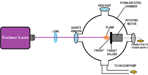
Fig 1: Schematic diagram of experimental setup.
Afterward transitory through a plane-convex lens with a focal length of 50 cm, the incident laser beam was directed vertically to the mark surface inside the chamber. The target samples were subjected with fixed 2250 number of pulses for five distinct laser fluences of 3.8, 4.3, 4.7, 5.1 and 5.5 J/cm2. A Scanning Electron Microscope (SEM) (JEOL-JSM-6480LV) was used to examine the surface morphology of the irradiation targets. An X-ray diffractometer (X’Pert PRO MPD) was used to analyze the phase and crystallographic structure of the exposed targets.
- Surface Analysis
3.1 Surface morphology
Figure 4.35 shows scanning electron micrographs taken in an oxygen atmosphere of (a) a unirradiated Aluminum-copper alloy and (b) an Aluminum-copper alloy that has been laser-irradiated at many stages. Figure 4.35 (b) displays a small number of LIPSS with a cyclicity of 2.5 µm at the minimum rate of 3.8 J/cm2. Raising the fluence to 4.3 J/cm2 has raised the ripples’ periodicity to 2.9 µm. The significant amounts of energy deposition, material deformation, and imprinting are responsible for this rising trend of periodicity with increasing laser fluence [5]. Figure 4.35 (d) shows that with a further increase in fluence up to 4.7 J/cm, gentle laser-induced episodic exterior structures (LIPSS) with a cyclicity of 2.5 µ m grow. Distinct periodic structures and diffused structures may be shielded by oxygen gas, which causes a decrease in periodicity. This phenomenon is associated with re-solidification, large-scale melting, and mergers [6], [7, 8]. Several events, including hydrodynamic Kelvin-Helmholtz instabilities that occur during surface modification, are associated with the development of laser-induced periodic surface structures (LIPSS) [9]. The expansion under pressure of the heated aluminum-copper alloy causes it to become a fluid, which in turn generates wind. After being splashed, re- solidified, icy, and impressed onto the ablated surface of the target material, micro-sized waves are produced [10]. It is possible that thermo-capillary waves have an effect on the formation of periodic formations created by lasers [11]. As the stage is raised to 5.1 J/cm2, Figure 4.35 (e) shows a distinct periodic structure with a periodicity of 2.9 µm. At a supreme fluence of 5.5 J/cm2, the cyclicity of the assemblies has diminished to a level of 2.5 µm, as seen in Figure 4.35 (f). The cyclicity of the waves rises as the laser fluence is raised from 3.8 to 4.3 J/cm2. For these fluences, it was shown that the material becomes softer when periodicity is increased, leading to a decrease in yield strength [12]. As the fluence reaches 4.7 J/cm2, the temperature gradient and solidification rate cause dense periodic patterns to form, which in turn boost the material’s toughness owing to the microstructure’s fineness [13].
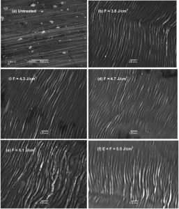
Fig 2: SEM micrographs showing how different steps affected the surface morphology of current work compound in an oxygen gas environment.
A rise in stage near to 5.1 J/cm2 reasons a decrease in ripple density as the spacing between periodic surface structures grows; a decrease in periodicity at 5.5 J/cm2, leading to a change in the hardness of the material, causes an increase in ripple density.
3.2. XRD analysis
It is possible to find out a material’s structure, phase identification, dislocation density, and energy deposited using the XRD method. The figure 3 displays the X-ray Diffraction patterns of (a) an Aluminum-copper alloy that has been unearthed and (b) an Aluminum-copper alloy that has been exposed to laser radiation, in an oxygen atmosphere.
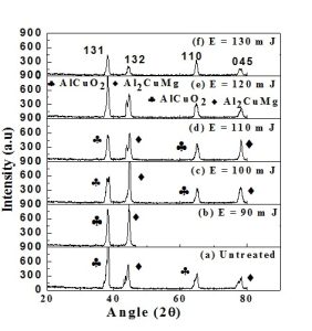
Fig 3: Different stages were used to generate patterns of current work alloy in an oxygen gas atmosphere.
Angles with stages (Al2MgO4, Cu4O3, AlMgO4, Al2MgO3) are detected for the un-irradiated mark diffraction points. With a little angular shift of 10 degrees, the diffraction peaks at angles 390, 450, 660, and 790 for the laser-irradiated target correspond to the (131), (132), (110) and (045) planes, respectively. Tensile stresses are indicated by peak moving toward the lower angular position, while compressive stresses are indicated by peak shifting toward the higher angular position [14]. Additionally, it has been noted that following laser ir-radiation of this alloy, new points (Al2CuMg, AlCuO2) have formed. The development of these stages is ascribed to the dispersal of oxygen and the fast degree of hotness and coldness brought on by laser irradiation. The creation of new phases is also caused by the melting, re-solidification, and redeposition processes brought on by laser ir-radiation [15]. The growth of scattered new-stage particles relative to the unique phase during the precipitation hardening process may boost the Aluminum-copper alloy’s hardness and tensile strength. For different fluences, the Aluminum-copper alloy specimen’s peak intensity, crystallite size, and dislocation density exhibit aberrant behavior respectively. Changes in crystallite size following laser irradiation and variations in laser-induced strain on the surface are related to variations in the average peak intensity in an oxygen environment, which can fluctuate. This alteration in remaining strain is linked to the fluctuation in lattice distortion and d-spacing. Differences in cooling and temperature conditions between the interstitial diffusion and surface layers, as well as the thermal expansion coefficient and differences in interatomic distances, contribute to the variation in lattice distortion [16] [17].
Crystallite Size (D) = 0.9λ / FWHMcos θ (1)
The displacement concentration is measured by means of next method [18]
Dislocation Density = 1/(Crystallite Size)2 (2)
Figure 3 shows the size fluctuation of crystallites and figure 4 shows the density of dislocations at different laser fluences. Figures 2 and 3 show that, at low fluences (3.8 J/cm to 4.3 J/cm), both the peak intensity and the size of the crystallites grow. As shown in figures 2 and 3, the intensification of X-ray diffraction from the target following laser ablation and crystal development brought on by atomic diffusion across grain boundaries are responsible for this rise in peak intensity and crystal size. [19, 20]. In this case, the solute oxygen atoms separate on the target surface and diffuse across the grain boundaries as a result of increased heat generation and energy deposition. The increased concentration and diffusion of oxygen gas into the material is the cause of this increase in peak intensity for the plane (132) of phase Al2CuMg [21].
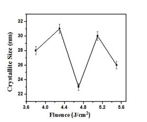
Fig 4: The change in the size of the crystallites produced current work alloy in an oxygen gas atmosphere at different stages at a recurrence degree of 30 Hz.
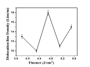
Fig 5: Stages were used to irradiate an oxygen gas environment and examine the change in dislocation density of an Aluminum-copper alloy.
Rapid melting and cooling, re-crystallization, and re-solidification follow laser irradiation, leading to a additional upsurge in fluence equal to 4.7 Joule/ square centimeter. The drop in highest power is caused by the disintegration of larger grains into smaller ones [22]. The peak intensity decreases as a result of widening as the laser fluence is raised to 5.1 Joule/ square centimeter, while the crystal size increases as a result of the recrystallization and reorganization of the material. When the stage is raised to a extreme of 5.5 J/cm2, the peak level is once again diminished. It is possible to associate a shift in peak intensity with a shift in d-spacing in this case. Changes in distortion of lattices and residual stresses are caused by variations in d-spacing. The transformation of tensile tensions into compressive stresses is achieved by raising the laser fluence.
Laser energy deposition causes lattice distortion and thermal shock waves, which in turn cause these stresses to develop [23]. Tensile strains and a temperature gradient are initially produced by the laser-induced shock waves. Tensile tensions relax and change into compressive stresses when fluence increases because of improved oxygen diffusion, which results in a reduction in crystallite size. [24]. Consequently, there is a trend toward decreasing peak intensities and increasing crystallite sizes, which suggests an unusual pattern of behavior. Similarly, for fluences between 3.8 and 4.3 J/cm2, dislocation density decreases, but it rises for fluences over 4.7 J/cm2. As the concentration of defects in the lattice sites increases, the dislocation density also increases. With more accurate measurements of work hardening, the crystal lattice’s dislocation line density rises. The energy received during laser-matter interaction causes deformation or vibration, which in turn creates new dislocation faults and makes pre-existing ones more mobile [25]. Therefore, an unexpected trend is suggested by both the size of the crystallites and the density of dislocations. Thermal stresses, lattice flaws, and recovery mechanisms can all play a role in this shift in density of dislocations and crystallite size. [26, 27].
Conclusion
In an oxygen environment, the exterior and structure characteristics of the Aluminum-copper alloy are highly connected after exposure to lasers with varied fluences. In comparison to nitrogen and vacuum, oxygen also significantly alters the exterior and structural characteristics of Aluminum-copper alloy. As the occurrence of the ripples is increased, the material softens and its yield strength decreases within the intensity range of 3.8 to 4.3 Joule per centimeter [12]. At fluences of up to 4.7 Joule per centimeter, solid episodic assemblies form as a result of an improved thermal gradient and solidification rate; these, in turn, boost the material’s mechanical characteristics and hardness as a result of its microstructure’s fineness [13]. Variations in dislocated density, the arrangement of crystals, structural defects, thermal conductivity, ionization capacity, and other variables might impact the hardness of a material when subjected to an oxygen irradiation surroundings. The mechanical characteristics and hardness of the material are also greatly affected by the type and pressure of the surrounding gas. Increasing the laser fluence may enhance the diffusion of O2 atoms along the grain boundaries, which in turn increases the micro hardness of the material [28]. A decrease in crystallite size and an increase in hardness are caused by the transformation of tensile stresses into compressive stresses, which occur as a result of the diffusion of these oxygen atoms.
Author’s Contribution: D.Y. & S.B., Conceived the idea; D.Y. & S.B. Designed the simulated work or acquisition of data; D.Y., Executed simulated work, data analysis or analysis and interpretation of data and wrote the basic draft; D.Y. & S.B., Did the language and grammatical edits or Critical revision
Funding: The publication of this article was funded by no one.
Conflicts of Interest: The authors declare no conflict of interest.
Acknowledgment: The authors would like to thank the advisors who advised for assistance with the collection of data.
References
[1] M. N. A. Grabowski, J. Sleziona, , “Laser cutting of an AlSi alloy composites: theory and experiments,” Journal of Achievements in Materials and Manufacturing Engineering, vol. 17, pp. 61-64, 2006.
[2] E. P. T. Tarasova, ” Influence of laser radiation on structure and properties of steels and alloys,” New achievements in materials and environmental sciences, vol. 416, 2013.
[3] S. L. L. Sexton, G. Byrne, A. Kennedy, , ” Laser cladding of aerospace materials,” J. Mater. Process. Technol, vol. 122, pp. 63-68, 2002.
[4] D. E. L. W.F. Miao, “Precipitation hardening in Aluminium alloy 6022,” Scripta Materialia, vol. 40, pp. 873–878, 1999.
[5] J. J. Yu, Y.F. Lu, “Laser-induced ripple structures on Ni–P substrates,” App. Surf. Sci., vol. 148, pp. 248-252, 1999.
[6] F. Z. M. Huang, N.Xu, Z. Xu, “The morphological and optical characteristics of femtosecond laser-induced large-area micro/nanostructures on GaAs, Si, and brass.,” Optics Express, vol. 18, pp. A601-A619, 2010.
[7] W.W.Duley, UV Laser: Effects and Applications in Materials Science. New York: Cambridge University Press, 1996.
[8] J. P. D.C. Emmony, J. H. Toyer, L. J. Willism, , “The topography of laser irradiated germanium,” Journal of Physics D: Applied Physics, vol. 8, pp. 1472-1479, 1975.
[9] L. K. Ang, Y. Y. Lau, R. M. Gilgenbach, H. L. Spindler, J. S. Lash, “Surface instability of multipulse laser ablation on a metallic target,” J. App. Phy., vol. 83, pp. 4466-4471, 1998.
[10] Y. F. Lu, J. J. Yu, W. K. Choi, “Theoretical analysis of laser-induced periodic structures at silicondioxide/ silicon and silicon-dioxide/aluminum interfaces,” Appl. Phys. Lett., vol. 71, p. 3439, 1997.
[11] X. C. Wang, et al., “Femtosecond pulsed laser-induced periodic surface structures on GaN/sapphire,” Applied Surface Science, vol. 252, pp. 1492-1497, 2005.
[12] J. A. E. P.G. Sanders, J.R. Weertman, , “Elastic and tensile behavior of nanocrystalline copper and palladium,” Acta Mater, vol. 45, pp. 4019- 4025, 1997.
[13] N. C. M. A. Pinto, M.C.F. Lerardi, A. Garcia, , “Microstructural and hardness investigation of an Al-Cu alloy processed by laser surface melting,” Materials Characterization, , vol. 50, pp. 249-253, 2003.
[14] D. P. L. Gaoren, J. H.Li, R.Y.Lin, “Nano Cr interlayered CrN coatings on steels,” Tsinghua Science and Technology, vol. 10, pp. 690-968, 2005.
[15] R. A. Umm-i-Kalsoom, N. Ali, I. A. Khan, S. Saleem, U. Ikhlaq, N. Khan, “Effect of Power and Nitrogen Content on the Deposition of CrN Films by Using Pulsed DC Magnetron Sputtering plasma,” Plasma Sci. & Technol. , vol. 15, pp. 666-672., 2013.
[16] D. Choi, R. J. Shinavski, W. S. Steffier, S. M. Spearing, “Residual stress in thick low-pressure chemical-vapor deposited polycrystalline SiC coatings on Si substrates,” J. Appl. Phys., vol. 97, pp. 074904-074909, 2005.
[17] S. R. S. B. D. Cullity, Elements of X-ray diffraction, 3rd ed.: Prentice Hall, 2001.
[18] S. Venkatachalam, K. R.T. Rajendra, D. Mangalaraj, S.K. Narayandass, K. Kim, and Y. Junsin, “Optoelectronic properties of Zn 0.52 Se 0.48/ Si Schottky diodes,” Sol. Elect., vol. 48, pp. 2219-2223, 2004.
[19] M. K.-u.-R. Latif. A, M. S.Rafiq, K. A. Bhatti, “Surface morphologic and structural analysis of IR irradiated silver,” Physica B, Consened matter, vol. 406, pp. 1713–1716, 2011.
[20] M. K.-u.-R. Latif. A, M. S.Rafiq, K. A. Bhatti, “IR and UV irradiations on ion bombarded poly crystalline silver,” Physica B, Condensed matter, vol. 405, pp. 4250-4255, 2010.
[21] N. F. K. Mahmood, I. M. Ghauri, N. Afzal, Y. Idrees, F. E. Mubarik, and . “Effects of laser irradiation on the mechanical response of polycrystalline titanium, ,” Phys. Scr, vol. 82, 2010.
[22] M. K. R. M. S. Rafique, T. Firdos, K. Aslam, M. S. Anwar, M. Imran, H. Latif, , “XRD and SEM analysis of a laser-irradiated Cadmium,,” Laser Phys, vol. 17, pp. 1138-1145, 2007.
[23] I. A. Khan, M. Hassan, R. Ahmad, A. Qayyum, G. Murtaza, M. Zakaullah, R.S. Rawat, , “Nitridation of zirconium using energetic ions from plasma focus device,” Thin Solid Films, vol. 516, pp. 8255–8263, 1 October 2008 2008.
[24] V. N. Gurarie, P.H.Otsuka, D.N. Jamieson, S. Prawe, “Crack-arresting compression layers produced by ion implantation,” Nuclear Instruments and Methods in Physics Research B, vol. 242, pp. 421–423, January 2006 2006.
[25] M. Z. B. M.K. Rahman, A. Samuel, K. Siraj, “Investigation of laser irradiation effects on the hardness of Al 5086 alloy under different conditions,” Vacuum, vol. 85, pp. 474 – 479, 2010.
[26] D. B. D. Hull, Introduction to dislocations. Oxford: Butterworth-Heinemann: Jordon Hill, 2001.
[27] W. F. Smith, Principles of Material Sciences and Engineering: Mc Graw Hill, 1990.
[28] J. W. Seok, N.M. Jadeed, R. Y. Lin, , Surface & Coatings Technology, vol. 138, pp. 14-22, 2001.